 English
  Español
(305) 442-0066
Coral Gables Office
Hialeah Office
North Miami Office
Hialeah Office
North Miami Office
Keratoconus is a condition where the cornea progressively thins and bulges like a cone. Changing the shape of the cornea makes the vision blurry and distorted. This can make daily tasks like reading and driving difficult to perform.
Keratoconus appears to have a genetic component and runs in families. It is also associated with allergies and excessive eye rubbing. Keratoconus often first presents in the teenage years and usually slowly progresses over a 10-20 year period. Keratoconus usually takes years to go from early- to late-stage. For some people, though, it can get worse quickly. The cornea can swell suddenly and start to scar. When the cornea has scar tissue, it loses its smoothness and becomes less clear. As a result, vision grows even more distorted and blurry.
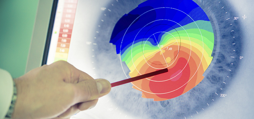
Symptoms of keratoconus include blurred or distorted vision, increase in the amount nearsightedness or astigmatism, inability to wear contact lenses. In order to diagnose keratoconus your doctor can perform topographic and tomographic images of the shape and curvature of the front and back surfaces of the cornea.
In very mild cases of keratoconus patients can usually achieve their desired vision with the help of glasses or soft contact lenses. As keratoconus progresses hard contact lenses may be utilized to restore a more normal shape and curvature to the cornea to help improve vision. If there is evidence that keratoconus is progressing by comparing topographic/tomographic images at two different time points, then corneal collagen crosslinking may be recommended. The cornea is made up of collagen fibrils which are weakened in keratoconus. If we saturate the cornea with riboflavin (Vitamin B2) drops and then expose it to UV light the collagen fibrils are able to form crosslinks between each other. This in turn strengthens the collagen fibrils and puts a stop to the progressive bulging of the cornea.
In more advanced cases where there has been significant scarring and all the previous treatments mentioned above are not effective corneal transplantation surgery may be recommended. (see Deep Anterior Lamellar Keratoplasty and Penetrating Keratoplasty sections).

2441 SW 37th Avenue
Miami, FL 33145
(305) 442-0066
202 E 49th Street
Hialeah, FL 33013
(305) 442-0066
2050 NE 163rd Street North
Miami Beach, FL 33162
Direct Line (786) 708-8372