 English
  Español
(305) 442-0066
Coral Gables Office
Hialeah Office
North Miami Office
Hialeah Office
North Miami Office
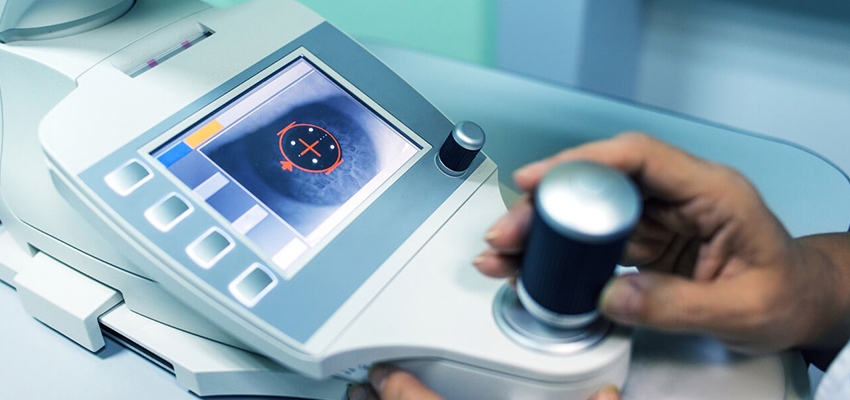
Photorefractive Keratectomy (PRK) uses a laser to sculpt the surface of the cornea by firing the laser directly into the corneal surface. This contrasts with LASIK, which removes deeper tissue within the cornea, under a corneal flap. In PRK, instead of creating a corneal flap, the surgeon scrapes the superficial corneal layer called epithelium over the treatment area. The second step of PRK is identical to LASIK: an excimer laser is used to remove stromal tissue and reshape the underlying corneal. In PRK, when more than 60 microns of tissue are removed an anti-scarring agent such as Mitomycin-C is used to allow the cornea to be crystal clear postoperatively.
PRK is useful for treating low to moderate levels of myopia, hyperopia and/or astigmatism. It is often the laser vision correction procedure of choice for people with thinner corneas, borderline tear secretion and for individuals who may have certain anterior corneal dystrophies, superficial corneal scars, or recurrent corneal erosions.
After the laser ablation, a soft contact lens is placed over the eye as a bandage while the corneal epithelium grows back in place, which usually takes about 4 to 5 days. During this period, the patient usually experiences mild to marked discomfort and the vision has variable degrees of blurriness. Because of the need for epithelial healing in PRK, it can take several weeks before vision is clear and stable after the procedure.
Even though PRK has more post-operative discomfort and longer healing and visual recovery time than LASIK, it is the procedure of choice in certain cases such as thin corneas, borderline tear secretion and borderline topography images. In all these cases, PRK offers significant advantages over LASIK. In general, the final visual outcome is similar in both techniques.

2441 SW 37th Avenue
Miami, FL 33145
(305) 442-0066
202 E 49th Street
Hialeah, FL 33013
(305) 442-0066
2050 NE 163rd Street North
Miami Beach, FL 33162
Direct Line (786) 708-8372